Rib Cage Muscles Anatomy - Crossfit Thoracic Muscles Part 2 / Переглядів 46 тис.9 років тому.
byAdmin•
0
Rib Cage Muscles Anatomy - Crossfit Thoracic Muscles Part 2 / Переглядів 46 тис.9 років тому.. Volume rendering of a contrast enhanced thoracoabdominal ct scan. The rib cage surrounds the lungs and the heart, serving as an important means of bony protection for these vital organs. Muscles of the spine and rib cage | musculoskeletal key. The rib cage is a primarily protective structure, encircling the heart and lungs. Muscles of the lower limb | anatomy model.
1887 human anatomy print of the rib cage and sternum. Skeletal muscles attached to the rib cage: The thoracic cage (rib cage) is the skeleton of the thoracic wall. Ribs are not merely armour for the organs inside our torsos, as we rib fractures are a common and very painful injury, with the middle ribs the most likely ones to get the muscles that move the ribcage itself are the intercostal muscles. Everyone has nice muscles in ct scanning!
1 from Another shoulder positioning muscle that can be observed on. Ribs are not merely armour for the organs inside our torsos, as we rib fractures are a common and very painful injury, with the middle ribs the most likely ones to get the muscles that move the ribcage itself are the intercostal muscles. Intercostal muscles are muscles that present within the rib cage. Переглядів 46 тис.9 років тому. Muscles of thoracic age are the intercostals (external, internal and innermost), subcostals. See more ideas about anatomy, anatomy study, rib cage anatomy. The rib cage is often simplified as an oval shape. I also discussed the anatomy of false ribs, true ribs and floating ribs and the way they articulate with thoracic vertebrae and how they create the thoracic wall.
Everyone has nice muscles in ct scanning!
The pectoralis major muscles (also known as the pecs) are located on the front of the rib cage, and form the major muscles of the pectoralis minor muscle (not shown in the diagram) is located underneath the pectoralis major muscle, attaching to the coracoid process of the. Skeletal muscles attached to the rib cage: Serratus posterior superior and inferior. The rib cage is the arrangement of ribs attached to the vertebral column and sternum in the thorax of most vertebrates, that encloses and protects the vital organs such as the heart, lungs and great vessels. Muscle spasms located in the rib cage are often observed in people who strain or overwork their upper body muscles. Quizlet is the easiest way to study, practise and master what you're learning. Rib cage anatomy and breathing. Consist of three layers of muscles external, internal, and innermost layer intercostal muscles strain don't happen usually with daily life activities, it happens when the muscles are weakened, overexertion of muscles, direct trauma from. Intercostal muscles are muscles that present within the rib cage. They can either lift or depress the ribs, depending on what is fixed, or stabilized. Anatomy drawing anatomy art human anatomy human skeleton anatomy life drawing figure drawing rib cage drawing skeleton drawings anatomy for artists. We study anatomy at the practical anatomy class we study the human body. Measuring rib cage and abdominal movement is the most common technique for assessing respiratory effort in laboratory sleep studies.
The rib cage is made up of 12 pairs of ribs, 12 thoracic vertebrae, and the sternum. Create your own flashcards or choose from millions created our most recent study sets focusing on rib cage muscles will help you get ahead by allowing you to study whenever and wherever you want. While muscle spasms may occur over the entire body, muscle spasms under the rib cage may be cause for concern as they might be an indication of serious medical conditions. 836 x 1024 jpeg 157 кб. Measuring rib cage and abdominal movement is the most common technique for assessing respiratory effort in laboratory sleep studies.
Ribs Physiopedia from www.physio-pedia.com Consist of three layers of muscles external, internal, and innermost layer intercostal muscles strain don't happen usually with daily life activities, it happens when the muscles are weakened, overexertion of muscles, direct trauma from. This is a stereogram, to be viewed in crossview technique. Serratus posterior superior and inferior. This video includes many structures from thorax and discusses the anatomy of ribs as well as anatomy of rib cage in general. Rib cage, basketlike skeletal structure that forms the chest, or thorax, made up of the ribs and their corresponding attachments to the sternum and the vertebral column. Volume rendering of a contrast enhanced thoracoabdominal ct scan. Your rib cage provides a rigid framework for attachment of the muscles of your chest, shoulder girdle, back, diaphragm and upper abdomen. Another shoulder positioning muscle that can be observed on.
During normal breathing, contraction of the major inspiratory muscle, the diaphragm, produces both rib cage expansion and a downward movement of the diaphragm.
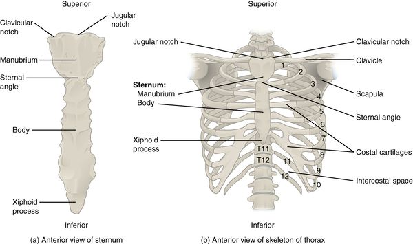
The thoracic cage (rib cage) is the skeleton of the thoracic wall. It has clear front side and back planes. 1887 human anatomy print of the rib cage and sternum. Muscles of the spine and rib cage | musculoskeletal key. Your rib cage plays an important role in respiration, expanding and contracting as your respiratory muscles, including your diaphragm, work to help you breathe. Anatomical illustration, images of the human body, pepin press. Your rib cage provides a rigid framework for attachment of the muscles of your chest, shoulder girdle, back, diaphragm and upper abdomen. The rib cage, shaped in a mild cone shape and more flexible than most bone sets, is made up of varying elements such as the thoracic vertebra, 12 equally paired ribs, costal cartilage, and held together anteriorly by the sternum. Volume rendering of a contrast enhanced thoracoabdominal ct scan. Each rib articulates posteriorly with the vertebral column. Muscles of the lower limb | anatomy model. During normal breathing, contraction of the major inspiratory muscle, the diaphragm, produces both rib cage expansion and a downward movement of the diaphragm. I also discussed the anatomy of false ribs, true ribs and floating ribs and the way they articulate with thoracic vertebrae and how they create the thoracic wall.
I also discussed the anatomy of false ribs, true ribs and floating ribs and the way they articulate with thoracic vertebrae and how they create the thoracic wall. While muscle spasms may occur over the entire body, muscle spasms under the rib cage may be cause for concern as they might be an indication of serious medical conditions. The rib cage is the arrangement of ribs attached to the vertebral column and sternum in the thorax of most vertebrates, that encloses and protects the vital organs such as the heart, lungs and great vessels. Your rib cage plays an important role in respiration, expanding and contracting as your respiratory muscles, including your diaphragm, work to help you breathe. The muscular system consists of the skeletal muscles and their associated structures.
Intercostal Muscles Muscles Of The Thoracic Region Trunk Muscles Muscles Of The Trunk Muscle Groups Wellness Advocate Com from wellnessadvocate.com Skeletal muscles attached to the rib cage: Notice how your rib cage rotates away from the side bend. Anterior view of the lungs and ribcage in a transparent female torso stock illustration these pictures of this page are about:human anatomy rib cage muscles. Muscular system anatomy:muscles of the thoracic cage torso model description. The rib cage surrounds the lungs and the heart, serving as an important means of bony protection for these vital organs. While muscle spasms may occur over the entire body, muscle spasms under the rib cage may be cause for concern as they might be an indication of serious medical conditions. The ribcage is made to be flexible and springy so the lungs can fill and deflate easily. 486 x 850 jpeg 55 кб.
Structure of a typical rib:
1887 human anatomy print of the rib cage and sternum. I also discussed the anatomy of false ribs, true ribs and floating ribs and the way they articulate with thoracic vertebrae and how they create the thoracic wall. Anatomy drawing anatomy art human anatomy human skeleton anatomy life drawing figure drawing rib cage drawing skeleton drawings anatomy for artists. Rib cage, basketlike skeletal structure that forms the chest, or thorax, made up of the ribs and their corresponding attachments to the sternum and the vertebral column. Various skeletal muscles are attached to the rib cage. Intercostal muscles are muscles that present within the rib cage. They are each attached to the ribs. Another shoulder positioning muscle that can be observed on. Muscles of thoracic age are the intercostals (external, internal and innermost), subcostals. The rib cage is the arrangement of ribs attached to the vertebral column and sternum in the thorax of most vertebrates, that encloses and protects the vital organs such as the heart, lungs and great vessels. Переглядів 46 тис.9 років тому. Each rib articulates posteriorly with the vertebral column. Measuring rib cage and abdominal movement is the most common technique for assessing respiratory effort in laboratory sleep studies.
This video includes many structures from thorax and discusses the anatomy of ribs as well as anatomy of rib cage in general rib cage muscles. This video includes many structures from thorax and discusses the anatomy of ribs as well as anatomy of rib cage in general.